This blog post is not an accusation of misconduct, and reflects my personal opinion.
Happy New Year! I started 2020 by scanning a set of papers from researchers at the Department of Pediatrics, West China Second University Hospital, Sichuan University, Chengdu, with a connection to the University of California in San Francisco (UCSF). These researchers use a pretty cruel baby rat model to investigate the effect of oxygen deprivation on the developing brain. I found that one out of five papers from this group appears to have image problems.
Like many of the other sets of papers with image concerns, this cluster started with a paper that I found during my scan of 20,000 papers in 40 journals, work that was published in 2016 in mBio.
The seed paper: Yi Qu et al., Mol Cell Biochem (2008)
One of the journals that I scanned for the mBio study was Molecular and Cellular Biochemistry, and one of the papers that I found in that journal was a 2008 paper by Yi Qu et al., with the title “Telomerase reconstitution contributes to resetting of circadian rhythm in fibroblasts“.
Figure 4 of that paper was of the typical low-resolution from the 2000s, but still struck me as having some repetitive patterns in two of the Western blot panels.

10.1007/s11010-008-9736-2, marked by me with red and blue lines to point out some repetitive features in two Western blot panels. Posted on PubPeer at https://pubpeer.com/publications/88B464FAC183D931498CE6A7452EDC .
I reported my concerns to the journal in October 2015, but the journal did not issue a correction or other explanation. After being patient for over 4 years, I posted my concerns on PubPeer two days ago.
Scanning more papers from the Dezhi Mu group
A couple of days ago I decided to scan more papers by this group, and focused – as I usually do – on papers published by the same first, Yi Qu, or last author, Dezhi Mu.
According to his ORCID page, Mu is the Assistant Dean of Sichuan University / West China Second University Hospital. A webpage from the Division of Neonatology from that hospital describes Mu as follows: “Professor MU Dezhi is academic leader. He is president of West China Second University Hospital of Sichuan University and vice chairman of the Subspecialty Group of Neonatology of Pediatric Society of Chinese Medical Association as well. Professor Mu is the first neonatal physician who holds the grant of the China National Funds for Distinguished Young Scientists medicine in China.”
Mu’s ORCID site also lists he is a Visiting Professor at the University of California San Francisco (UCSF) since 2005. A search on the UCSF website could not confirm if this affiliation is still ongoing, but Mu and his wife appear to own (or have owned) a house near San Francisco.
Update: In a 2013 post on Dr. Fang Zhouzi’s website XYS.org (in Chinese, English translation here), it was discussed that Dezhi Mu might have misrepresented his UCSF affiliation, which he claimed was an Associate Professor. A reader suggested he might have only been a temporary research assistant or postdoc at UCSF, based on the website of the research group at UCSF where he worked and his salary listings. This raises additional concerns and might mean that some of the papers discussed in this post might have been published under a false affiliation.
Yi Qu is listed on ORCID as a Professor in the Department of Pediatrics at the same West China Second University Hospital. She has a PhD in Biomedical Engineering and did a postdoc at the University of Hong Kong.
Mu’s research group has published about 150 PubMed-indexed papers, according to this search limiting for the Sichuan University affiliation. Excluding review papers, meta-analyses, cohort studies, and case reports, about 80 of these papers contained photos of Western blots or tissues.
Including the paper mentioned above, I found 18 of these 80 papers to contain possible inappropriate image duplications. That is roughly one out of five papers. These duplications were found either between panels in different papers, between panels within the same paper, or within the same photo.
Several of Mu’s papers are published with his UCSF affiliation and/or with his UCSF email address provided as the corresponding author contact information, although all of the work appears to have been conducted at Sichuan University.
For each of the 18 papers with image problems, I wrote a PubPeer post. You can find all of Mu’s papers flagged on PubPeer by this search. I also posted a list of all 18 at the end of this post, but here are some remarkable findings.
Hypoxia and Ischemia baby rat model
Mu’s earlier papers describe the Hypoxia/Ischemia rat model to study brain injury in neonates. This model involves exposing 10-day old rat babies to hypoxia, very low oxygen content. In this case they put the pups for 2.5 hours in a chamber with 8% O2. That is the oxygen level at 26,000 ft/8,000 m altitude, the equivalent of the highest mountains on earth, such as those in the Himalayas.
To add injury to insult, some of these pups were also operated on to tie off their right common carotid artery (the large blood vessel on the right side of the neck that supplies the head with oxygen-rich blood). The researchers closed off this artery with a silk (classy!) suture, which will completely block off the blood supply to one side of the head, a condition called ischemia. This condition would likely induce a massive stroke. Finally, a third set of pups only received a “sham” operation (in which they were operated but their artery was not tied). These animals served as a control group.

You should now be gasping for air, because these are horrible animal experiments. Just to clarify, I am not opposed to animal experiments per se. They are sometimes necessary to learn about ways to help fight or prevent human disease (in this case to learn about stroke or brain injuries in newborns). But I am very sad to see these types of experiments in papers with image concerns, because those duplications might indicate problems regarding the general quality of the experiments in these papers.
So let’s take a look at some of the problems I found in Mu’s Hypoxia/Ischemia papers.
Two papers by Lihua Li et al.
Sham or Hypoxia (H) or Hypoxia+Ischemia (HI)? It is not clear if you look at this photos, one of which was used in two different papers.

Yi Qu et al., Stroke (2009)
Let’s take a look at another paper from the Mu lab that uses the Hypoxia/Ischemia rat model. In Yi Qu et al., Stroke (2009), HI was induced in baby rats by ligating their neck artery and putting them in the Himalaya box. Then, the poor pups were treated with a short piece of DNA (called an oligonucleotide), to see if that could prevent cell death by blocking the expression of a protein called HGTD-P.
In Figure 3 and 4 of the Qu paper, the researchers analyze the HGTD-P expression in several differently-treated rat pups. Here is the legend to the abbreviations.
All of the photos below should look differently. A photo from a Sham control rat should not look identical to that of a “Hanks” (HI-induced treated with buffer) rat. Or a photo from a HI rat should not look like that of a HI rat treated with an oligonucleotide. All these rats were differently-treated individual animals, and although their tissues might look similar, their tissues should never identical.
But – guess what I found? You guessed it: more possible duplications.

Legend to the abbreviations:
- Sham: control rats (did not undergo Hypoxia or Ischemia)
- HI: rats who underwent the Hypoxia and Ischemia procedures
- Hanks: HI rats treated with buffer alone
- SE: HI rats treated with a “sense” oligonucleotide
- AS: HI rats treated with an “antisense” oligonucleotide.
Boxes of identical colors point attention to tissues that look much more similar than expected. Also note that the photos are stretched differently and/or shown with different color intensities, suggesting these were not simple “oops I inserted the wrong image” types of errors.
Yi Qu et al. Stroke (2011)
This 2011 Stroke paper by Yi Qu et al. deserves special attention, since it appears to have so many image problems. Here are some highlights, but you can find all of my concerns about this paper on PubPeer.
In this study, the researchers again did the Hypoxia/Ischemia treatment on 10-day old rat pups, and they studied the expression of an injury-related protein called TERT in astrocytes of the pups after HI treatment.
Figure 1C of this paper appears to show some repetitive features, marked by me with ellipses of the same color.

Posted on PubPeer at: https://pubpeer.com/publications/9E1ACF40812F2299A42AC199389BD2
Figure 4B of this paper also appears to show some repetitive features, marked by me with ellipses of the same color.

Posted on PubPeer at: https://pubpeer.com/publications/9E1ACF40812F2299A42AC199389BD2
Figure 4C of this paper also appears to show some repetitive features, marked by me with ellipses of the same color.

Posted on PubPeer at: https://pubpeer.com/publications/9E1ACF40812F2299A42AC199389BD2
Figure 5A of this paper also appears to show some repetitive features, marked by me with ellipses of the same color.

Posted on PubPeer at: https://pubpeer.com/publications/9E1ACF40812F2299A42AC199389BD2
This might become repetitive, but Figure 6B and 6C of this paper also appear to show some repetitive features, marked by me with ellipses of the same color. Note that I did not mark all of these repeats, just enough to make a point.

Posted on PubPeer at: https://pubpeer.com/publications/9E1ACF40812F2299A42AC199389BD2
In addition, PubPeer user Hoya Camphorifolia had some questions about Figure 3A.

https://pubpeer.com/publications/9E1ACF40812F2299A42AC199389BD2
Yi Qu et al. Neurobiology of Disease (2014)
In 2014, the Dezhi Mu lab still was tying off baby rat arteries and putting them in low-oxygen boxes. They must have used 100s of baby rats at this point. But the image duplications appear to continue. Here are some examples from images of concern from a Yi Qu et al. paper published in Neurobiology of Disease (2014) as pointed out by me on PubPeer.
In Figure 2A, the same gel lane appears to be visible twice. Note the presence of two dots and a slanted line above the band.


A bit of a stretch
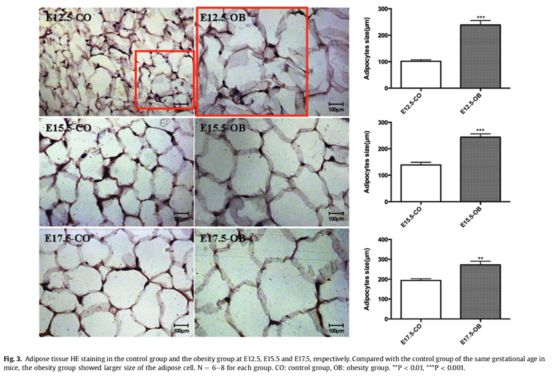
Stretching, flipping, or rotating images could be an indication that a figure was not just duplicated as the result of an honest error. Here are some examples where photos might have been stretched. Would you have spotted these?

In the example below, the obesity group (OB) showed larger fat cells than the control group (CO). Were the OB fat cells really larger? I found that to be a bit of a stretch.
Funding sources
Most of the papers that I found image problems in have been funded by the National Natural Science Foundation of China (multiple grants awarded to Yi Qu or Dezhi Mu) and the Outstanding Young Scientist Foundation of Sichuan Province, awarded to Yi Qu. Several other grants are mentioned as well, including from the China Medical Board of New York, the Ministry of Education of China, the Major State Basic Research Development Program, the Bureau of Scientific Technology of Sichuan Province, the Program for Changjiang Scholars and Innovative Research Team in University, the State Commission of Science Technology of China, the Grant of Clinical Discipline Program (Neonatology) from the Ministry of Health of China.
It is probably fair to say that the equivalent of millions of US dollars has been spent on these studies. But I am most sad about the hundreds of baby rats who died in agony for these papers with little progress for science.
Update 2: A Twitter user known to me has written a post on Weibo (Chinese social media site) about the research and PubPeer-flagged papers authored by Yi Qu and Dezhi Mu. They estimated that the combined Qu and Mu’s NNSF grant funds amount to a total of 7.42 million RMB, which is about 1 million US dollar. Here is a screenshot of their Weibo post, which as of today (Sat, Jan 4, 2020) has almost 50K views.

All 18 flagged papers from the Dezhi Mu group
Here is a table of all papers from Dezhi Mu et al. with concerns (n=18 as of now) with links to their PubPeer entries as well.
| Authors | Title | Citation | DOI | PubPeer link |
| Lihua Li , Yi Qu , Jinhui Li , Ying Xiong , Meng Mao , Dezhi Mu | Relationship between HIF-1α expression and neuronal apoptosis in neonatal rats with hypoxia–ischemia brain injury | BRAIN RESEARCH 1180 (2007) 133–139 | 10.1016/j.brainres.2007.08.059 | https://pubpeer.com/publications/5E553C4B253F3AB37837DDB05057DC |
| Yi Qu, Meng Mao, Xihong Li, Yanyou Liu, Jianmin Ding, Zhou Jiang, Chaomin Wan, Lin Zhang, Zhengrong Wang, Dezhi Mu | Telomerase reconstitution contributes to resetting of circadian rhythm in fibroblasts | Mol Cell Biochem (2008) 313:11–18 | 10.1007/s11010-008-9736-2 | https://pubpeer.com/publications/88B464FAC183D931498CE6A7452EDC |
| Lihua Li · Ying Xiong · Yi Qu · Meng Mao · Weiya Mu · Hua Wang · Dezhi Mu | The requirement of extracellular signal-related protein kinase pathway in the activation of hypoxia inducible factor 1a in the developing rat brain after hypoxia–ischemia | Acta Neuropathol (2008) 115:297–303 | 10.1007/s00401-008-0339-5 | https://pubpeer.com/publications/A0CCC03D7593A9F5C4D796A6D18145 |
| Xiaomei Sun , Hui Zhou , Xiaoli Luo , Shengfu Li , Dan Yu , Jiping Hua , Dezhi Mu , Meng Mao | Neuroprotection of brain-derived neurotrophic factor against hypoxic injury in vitro requires activation of extracellular signal-regulated kinase and phosphatidylinositol 3-kinase | Int. J. Devl Neuroscience 26 (2008) 363–370 | 10.1016/j.ijdevneu.2007.11.005 | https://pubpeer.com/publications/ED1F35595C9CE803CAC4E92E64E683 |
| Yi Qu , Meng Mao , Fengyan Zhao , Lin Zhang , Dezhi Mu | Proapoptotic Role of Human Growth and Transformation-Dependent Protein in the Developing Rat Brain After Hypoxia-Ischemia | Stroke. 2009;40:2843-2848 | 10.1161/STROKEAHA.109.553644 | https://pubpeer.com/publications/F4A8E58A6AEEFD761C030D58F0E766 |
| Yi Qu, Zhoujin Duan, Fengyan Zhao, Dapeng Wei, Jianbo Zhang, Binzhi Tang, Jiao Li, Chunlei Yang, Dezhi Mu | Telomerase Reverse Transcriptase Upregulation Attenuates Astrocyte Proliferation and Promotes Neuronal Survival in the Hypoxic–Ischemic Rat Brain | Stroke. 2011; 42:3542-3550 | 10.1161/STROKEAHA.111.626325 | https://pubpeer.com/publications/9E1ACF40812F2299A42AC199389BD2 |
| Hua Wang , Ying Xiong , Dezhi Mu | PirB restricts neuronal regeneration in developing rat brain following hypoxia-ischemia | MOLECULAR MEDICINE REPORTS 6: 339-344, 2012 | 10.3892/mmr.2012.907 | https://pubpeer.com/publications/033C258855A3328EFC0F6CAF086F98 |
| Yi Qu, Jinlin Wu, Dapeng Chen, Fengyan Zhao, Junyan Liu, Chunlei Yang, Dapeng Wei, Donna M. Ferriero, Dezhi Mu | MiR-139-5p inhibits HGTD-P and regulates neuronal apoptosis induced by hypoxia–ischemia in neonatal rats | Neurobiology of Disease 63 (2014) 184–193 | 10.1016/j.nbd.2013.11.023 | https://pubpeer.com/publications/67D613FA97B87C373F239931962126 |
| Yifei Li , Jie Fang , Yimin Hua , Chuan Wang , Dezhi Mu , Kaiyu Zhou | The Study of Fetal Rat Model of Intra-Amniotic Isoproterenol Injection Induced Heart Dysfunction and Phenotypic Switch of Contractile Proteins | BioMed Research International Volume 2014, Article ID 360687, 14 pages | 10.1155/2014/360687 | https://pubpeer.com/publications/DF2E0BC3E2927ECB9099F49E808D48 |
| Yi Qu, Xiang Huang , Zhiqing Li , Junyan Liu , Jinlin Wu , Dapeng Chen , Fengyan Zhao , Dezhi Mu | miR-199a-3p Inhibits Aurora Kinase A and Attenuates Prostate Cancer Growth | The American Journal of Pathology, Vol. 184, No. 5, May 2014 | 10.1016/j.ajpath.2014.01.017 | https://pubpeer.com/publications/97E5393475EBFF0025FE135040BA33 |
| Deyuan Li, Xihong Li, Jinlin Wu, Jinhui Li, Li Zhang, Tao Xiong, Jun Tang, Yi Qu, Dezhi Mu | Involvement of the JNK/FOXO3a/Bim Pathway in Neuronal Apoptosis after Hypoxic–Ischemic Brain Damage in Neonatal Rats | PLoS ONE 10(7): e0132998 | 10.1371/journal.pone.0132998 | https://pubpeer.com/publications/4841782C492CDFD2D16B68C9C19F98 |
| YunJie Su1, Yi Qu1,2, FengYan Zhao1,2, HuaFeng Li2,4, DeZhi Mu1,2,3 and XiHong Li | Regulation of autophagy by the nuclear factor κB signaling pathway in the | Journal of Neuroinflammation (2015) 12:116 | 10.1186/s12974-015-0336-2 | https://pubpeer.com/publications/490C9693DA13AD20460FDD08CD831B |
| Chuan Wang , Huaying Li , Chunyan Luo , Yifei Li , Yi Zhang , Ding Yun , Dezhi Mu , Kaiyu Zhou author has email , Yimin Hua | The effect of maternal obesity on the expression and functionality of placental P-glycoprotein: Implications in the individualized transplacental digoxin treatment for fetal heart failure | Placenta (2015) | 10.1016/j.placenta.2015.08.007 | https://pubpeer.com/publications/CDB819625AB397B2F60FE01C1AAADF |
| Yi Qu, Jing Shi, Ying Tang, Fengyan Zhao, Shiping Li, Junjie Meng, Jun Tang, Xuemei Lin, Xiaodong Peng, Dezhi Mu | MLKL inhibition attenuates hypoxia-ischemia induced neuronal damage in developing brain | Experimental Neurology 2016 | 10.1016/j.expneurol.2016.03.011 | https://pubpeer.com/publications/65A35A03A00E2D1EEC4837D1BB794C |
| Qianyun Cai , Jing Gan , Rong Luo , Yi Qu , Shiping Li , Chaomin Wan , Dezhi Mu | The role of necroptosis in status epilepticus-induced brain injury in juvenile rat | Epilepsy & Behavior 75 (2017) 134–142 | 10.1016/j.yebeh.2017.05.025 | https://pubpeer.com/publications/5D5B57AA93F3677A1639FB6AF1666D |
| Yi Qu , Jun Tang , Huiqing Wang , Shiping Li , Fengyan Zhao , Li Zhang , Q Richard Lu , Dezhi Mu | RIPK3 interactions with MLKL and CaMKII mediate oligodendrocytes death in the developing brain | Cell Death and Disease (2017) 8, e2629 | 10.1038/cddis.2017.54 | https://pubpeer.com/publications/F926FAB71D30C724D1D02E67A1F0C1 |
| Fengyan Zhao , Yi Qu , Huiqing Wang , Lan Huang , Jianghu Zhu , Shiping Li , Yu Tong , Li Zhang , Jiao Li , Dezhi Mu | The effect of miR-30d on apoptosis and autophagy in cultured astrocytes under oxygen-glucose deprivation | Brain Research (2017) | 10.1016/j.brainres.2017.06.011 | https://pubpeer.com/publications/72A9FA9DF3C3FBBDD0B1B6AC7E8025 |
| Laifeng Ren , Ming Zeng , Zizhi Tang , Mingyuan Li , Xiaojun Wang , Yang Xu , Yuding Weng , Xiaobo Wang , Huan Wang , Liandi Guo , Bing Zuo , Xin Wang , Si Wang , Jiangyan Lou , Yaxiong Tang , Dezhi Mu author has email , Ning Zheng , Xianhui Wu , Junhong Han , Antony M. Carr , Penelope Jeggo, Cong Liu | The Antiresection Activity of the X Protein Encoded by Hepatitis Virus B | Hepatology, Vol. 69, No. 6, 2019 | 10.1002/hep.30571 | https://pubpeer.com/publications/D563A7F243F255F8889F5CEB849A59 |


you are nothing but an ugly bitch!
LikeLike
you are the best animal model because you just look like a fat female rat!
LikeLike
These remarks suggest you have a keenly focused scientific mind. They also suggest Dr. Bik has “struck gold” in these findings, since Yi Qu and Dezhi Mu’s defenders apparently have no recourse but to call her juvenile names.
LikeLike
good job.
LikeLike
I agree with Ban Hu!
LikeLike
Well done, Elisabeth!
Chinese research is suffering under a dictatorship with an extreme corruption level.
Their hierarchical culture does not making it better for true science.
There are many similar cases from China:
Qinglong Guo, China Pharmaceutical University
https://pubpeer.com/search?q=qinglong+guo
Jinmin Zhao, Guangxi Medical University
https://pubpeer.com/search?q=Jinmin+Zhao
Cao Xuetao, Nankai University
https://pubpeer.com/search?q=Xuetao+Cao
Xu Hanmei, China Pharmaceutical University
https://pubpeer.com/search?q=xu+hanmei
Zhangjian Huang, China Pharmaceutical University
https://pubpeer.com/search?q=Zhangjian+Huang
Conclusion so far: Almost no retractions (only by Journal of Biological Chemistry), a string of corrections
and business as usual for the PIs… Real science R.I.P
LikeLike
Wow impressive analysis.
Thank you for your service to the public.
LikeLike
Thanks for your work on this disturbing matter
LikeLike