
Surgisphere is a company that specializes in the analysis of clinical data. It provided the large datasets on COVID-19 patients that formed the basis of two papers recently published in The Lancet and The New England Journal of Medicine, both of which were retracted on June 4th. Suspicions had been raised about the validity of the data, provided by Surgisphere founder Sapan S. Desai (archived ResearchGate page) who was an author on both papers.
But that might not be the only concern about Desai’s work. Here I will discuss a paper from his PhD project at the University of Illinois at Chicago that appears to have some serious image problems.

Sapan Desai, Surgisphere, and COVID-19 data
Sapan S. Desai is a vascular surgeon who did his MD/PhD at the University of Illinois at Chicago, specializing in Anatomy and Cell Biology. According to his (archived) ResearchGate page , he graduated in 2006, and then worked at several hospitals as a surgeon. His most recent hospital appointment was at Northwest Community Hospital in Illinois, where he resigned earlier in 2020. This article in the Scientist, however, revealed that Desai was named in several medical malpractice lawsuits, which he claims were unfounded.
Desai founded Surgisphere in 2008. The company’s (archived) website reads “When Dr. Sapan Desai founded Surgisphere Corporation, the mission was simple: to harness the power of data analytics and improve the lives of as many people as possible.”. Its values include “Public Service, Scientific Scholarship, Meaningful Impact, and Pursuit of the Truth”.

The company was not very well-known, but became suddenly in May 2020 after it provided a large dataset from almost 100,000 COVID-19 patients treated at 671 hospitals across the world. This enormous dataset appeared to show that treatment of COVID-19 infection with hydroxychloroquine or chloroquine, either with or without antibiotics, did not help patients to get better. In fact, it showed that each of these treatments resulted in a higher risk of death and more heart problems.
The data was published in May 22, 2020 in The Lancet, with Mandeep R Mehra as the first author, Desai as the second author. Frank Ruschitzka and Amit N Patel were listed as authors as well.
A related dataset, also provided by Desai / Surgisphere, obtained from 8910 COVID-19 patients and 169 hospitals had been published on May 1st, 2020 in The New England Journal of Medicine. That paper showed that underlying heart disease was associated with an increased risk of in-hospital death. Among the five authors on this paper, Mehra was again the first author, and Desai the second.
Retraction of the Lancet and NEJM papers
The Lancet and NEJM are two top journals in the medical field, and the potential of Hydroxychloroquine to treat COVID-19 patients is very controversial, so both Mehra et al. papers were widely discussed online. However, immediately after publication, many questions were raised about the validity of these datasets. There were issues with the statistical analysis, ethics approval, surprisingly similar data across different countries, and questions about how such a large dataset could be obtained in a paper with only 4 authors.
Surgisphere did not want to share their dataset – not a good sign.
On May 28, an Open Letter co-signed by 182 scientists led by statistician Dr. James Watson was published that openly questioned the data validity and analysis in these papers.
On June 4th, within an hour from each other, both Lancet and NEJM papers were retracted at the requests of some – but not all – authors. Surgisphere founder Sapan S. Desai did not agree with the Lancet retraction.
More concerns about Desai’s older work
Twitter follower Michael Brown, an astronomer in Melbourne, Australia, suggested it might be a good idea to look at other papers by Sapan Desai, which I did. After scanning several of his papers, most of which looked fine or did not contain photos, I ended up with a paper from his PhD period at the University of Illinois in Chicago.
Comparative Morphology of Rodent Vestibular Periphery. II. Cristae Ampullares – Sapan S. Desai, Hussain Ali, and Anna Lysakowski – Journal of Neurophysiology (2005), doi: 10.1152/jn.00747.2003
Figure 4 in this paper contained a collage of 9 tissue sections obtained from different rodent species. The crista ampullaris is part of the inner ear, a small region containing hair cells that can sense rotation.

Melba Toast
The tissue sections in Figure 4 look remarkably similar to pieces of Melba Toast. And just so you can impress your friends at the next party (which might still be far in the future), Melba Toast is named after Dame Nellie Melba, an Australian opera singer, whose real name was Helen Porter Mitchell.
Of course, the slices in Desai’s paper are not really pieces of toast, but it is hard to think of anything else.
Repetitive features
Not only did Figure 4 made me hungry for a late-night snack, it also showed a lot of repetitive features. Biological tissues are not expected to contain repeated areas, because every cell looks a bit different, similar to rocks or faces. So this was unexpected.
Here is a close-up look at the Tree Squirrel piece of toast:
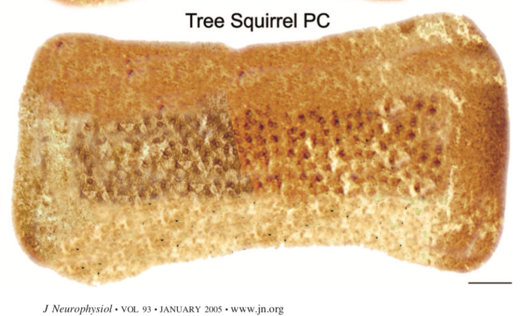
I first noticed some potential repeats at the bottom part of this image. You see a slightly darker, triangular piece of tissue with a small black dot in the middle. That piece appears to be visible multiple times. At the top, there are some brown spots that look very similar to each other. After spending quite some time on this image, I marked all areas that looked similar to each other with boxes of the same color.
Here is my marked version:

But the Tree Squirrel piece of toast was not the only one that showed such repeats. The whole party tray appeared to have these repetitive areas.

Concerns
As usual, this blog post is not an accusation of science misconduct.
There might be a perfectly acceptable biological explanation that could cause pieces of Melba Toast rodent cristae to contain repetitive areas. I just could not come up with one. There are also three authors on this paper, so it is not clear who prepared these photos.
Together with the questions raised about the Surgisphere dataset and Dr. Desai’s unwillingness to share the datasets described in the now retracted Lancet and NEJM papers, this figure raises additional concerns about this author.
I posted my concerns on PubPeer, with emails sent to the first and last authors. I am looking forward to their response.
The work described in this paper was supported by grants from the National Institute of Deafness and Communication Disorders Grant and the National Aeronautics and Space Administration Grant, and a National Institute of Deafness and Communication Disorders Predoctoral NRSA Fellowship F30 DC-05451 to S. S. Desai.


I had the same idea and started looking at the tables in the partner paper: “Comparative Morphology of Rodent Vestibular Periphery. I. Saccular and Utricular Maculae” https://doi.org/10.1152/jn.00746.2003
I haven’t finished yet, but there is enough that is odd to make we want to continue.
Fig 1B and 1C overlap at a different focal plane, but it might be that they are supposed to – I don’t understand the text.
LikeLike
Yes, those images overlapped, but I have not flagged them because they appeared to show two different fields of the same tissue sample.
LikeLike
Why would someone do this? Did he ruin ALL of his sections or something??
LikeLike
Curious about the discontinuity of shading in the middle of the tree squirrel PC, which you didn’t flag. How did you interpret that? Thanks!
LikeLike
This sort of image manipulation is easy to do in Photoshop. You can use the clone stamp tool to directly copy bits of the image from one place to another; then, to get rid of too obvious repetitions, you would use the spot healing brush tool to blur the edges of the cloned sections. There are various options on the healing brush tool: content aware (which makes a semi-intelligent guess about what to put in); create texture (which mimics adjacent textures with some small changes); and proximity match (which does a less intelligent job than content aware). There are several slightly different versions of the healing brush tool, for larger or smaller areas, but they all do the same sort of thing, that is, replace bits of the image with a rough guess as to what might plausibly go there. Note that while I speak of the tool as if it were making up its own mind, of course, it’s just a sophisticated bit of math, and the results can sometimes be unpredictable. To achieve similar results to those in the above images would take time and care, that is, it’s unlikely to be an accidental result of image processing.
LikeLike