A stem cell research group at the University of Louisville, Kentucky — famous for apparently discovering an exciting new class of stem cells — could be facing new troubles.
Although the work of Mariusz Ratajczak was supported through large NIH and Vatican grants, no other lab could replicate his findings on very small embryonic-like stem cells (VSELs).
And now, 28 papers from the Ratajczak lab are listed on PubPeer for image duplication and textual similarity concerns.

March 2022 update: No misconduct found
The University of Louisville sent their final decision – dated July 29, 2021 – to me today – March 23, 2022 (shared here with permission).
“The University of Louisville found there was no research misconduct. The institution followed its established, thorough, and robust process and made no findings of research misconduct against Dr. Ratajczak associated with any of the allegations, including all the allegations that continue to persist publicly on the internet. ”
A great discovery backed by the Vatican
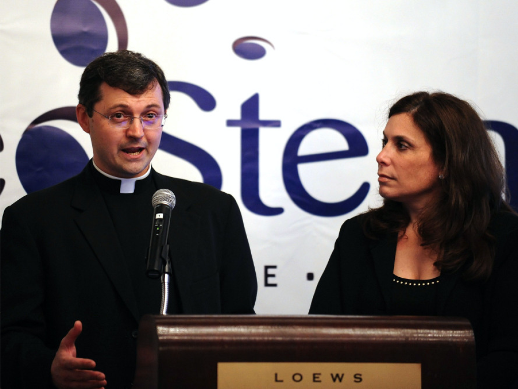
Mariusz Z. Ratajczak has received several awards for his work on stem cells. Around 2006, he discovered the presence of stem cells in the bone marrow of adult mice. These adult stem cells, known as VSELs, are primitive, undifferentiated cells with the potential to develop into many different kinds of specialized cells, such as heart, bone, brain, or pancreatic cells.
Ratajczak’s discovery of these VSELs was exciting because it could theoretically lead to all kinds of new treatment options. Patients with heart disease, burnt skin, diabetes, or neurodegenerative disorders, for example, might one day be cured simply by having their own bone marrow cells harvested and programmed to develop into specialized tissues that could restore their health.
This technique, harvesting bone marrow cells from adults, was in particular attractive to people who were opposed to using cells from human embryos. And when the company NeoStem began working with the patents filed by the Ratajczak lab in 2011, the Vatican took the unusual step of investing $1 million in the New York-based company.
NeoStem, now known as Caladrius Biosciences, also received a $1.2 million grant from the National Institute of Health for a project to repair bone defects using VSELs.
VSELs not confirmed by other labs
But other stem cell researchers were simply not convinced that VSELs can be programmed into differentiation, nor that VSELs were even real.
The lab of Irving Weissman at Stanford tried hard to replicate the Ratajczak findings, but was unable to detect these cells at all. In a 2013 paper in Stem Cell Reports, Weissman’s lab concluded that the existence of adult mouse VSELs was at best dubious.
“We tried as hard as we could to replicate the original published results using the methods described and were unable to detect these cells in either the bone marrow or the blood of laboratory mice.”, Dr. Weissman wrote in a Stanford press release.
An accompanying Cell Stem Cell profile article is also highly critical of the work by the Louisville lab. George Daley, back then the Director of Stem Cell Transplantation at Children’s Hospital, Boston said: “I find the work mystifying and lacking in rigor.’’
The headline of a July 2013 RawStory.com article on VSELs was even harsher.

Concerns in 26 papers
Not only have other labs found it impossible to reproduce Ratajczak’s work, his papers also appear to contain irregularities. In an investigation I carried out last year after receiving an anonymous tip, I confirmed image and textual similarity concerns in about two dozen papers from the Louisville lab. All concerns have been posted on PubPeer as of yesterday. There may be a couple more not yet listed on PubPeer, as I’m still going through the large volume of papers and similarities.
Duplicate figures across different papers
Sixteen of the 28 papers have concerns about image reuse. In most cases, it appears as though the images were representing the same experiments. This would be acceptable, so long as the paper acknowledged that the figure had been published before. However, in most of these cases the same figure appeared in multiple papers without citing older publications, while claiming that the results were novel.

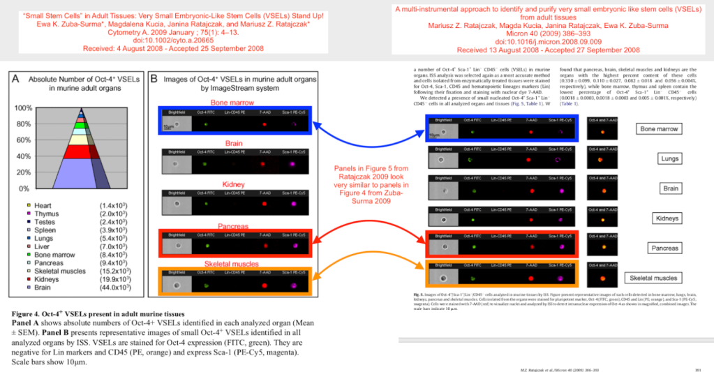
In one case, the same figure appears to have been published in four different papers by Ratajczak et al.

Overlapping images
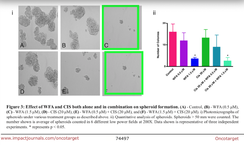
Some papers appear to contain figures with overlapping panels. In these cases, panels within the same figure seem to show areas of overlap, which I have highlighted using boxes of the same color. The panels are labeled differently, so they represent different experiments, and such an overlap would thus be highly unexpected.
Here is a figure from Wysoczynski et al. International Journal of Cancer (2010), DOI: 10.1002/ijc.24732:

An example from Kakar et al., Oncotarget (2017), DOI: 10.18632/oncotarget.20170:
And here is a figure from Libura et al., Blood (2002), DOI: 10.1182/blood-2002-01-0031 :

Duplications within photos
In some papers, the photos themselves appear to contain duplicated features, something that seems hard to explain as the result of an honest error.
Here is an example of such a potential duplication, from the Ratajczak lab’s highly cited Leukemia 2006 paper, DOI: 10.1038/sj.leu.2404171, cited 782 times:

Here is a paper by Shirvaikar et al, Stem Cells and Development (2011), that appears to show duplications within photos of gels and blots:

A 2018 PLOS ONE (2018) paper by Marlicz et al., DOI: 10.1371/journal.pone.0189337 may contain some irregularities in photos of gels:

A paper by Golan et al., Blood (2012), DOI: 10.1182/blood-2011-06-358614, appears to show highly similar — yet slightly different — flow cytometry panels:

And something is definitely not right in this photo from Huang et al., American Journal of Transplantation (2016), DOI: 10.1111/ajt.13511:

Text recycling?
Several papers by Ratajczak et al. appear to contain recycled text, wording that is highly similar in different papers. Text recycling, sometimes called self-plagiarism, is different from plagiarism, in which someone else’s text is copied and passed off as one’s own. Here, text is reused by the same researcher, but passed off as novel. Textual similarities are easy to prove using software such as SimTexter.
Here are some examples, with similarities indicated using highlighted text:


A list of all papers with PubPeer posts
This is a spreadsheet of the papers from the Ratajczak lab with image and textual similarities; 28 as of today. The list could grow, as I may confirm additional findings.


Dear Madam,
I feel greatly sorry to trouble you.
I am a resident in China and have a chance to study in a tertiary class hospital. As my major is radition oncology, I feel greatly excited at the first sceond. However, I found something wrong with my future tutor–https://pubpeer.com/search?q=Chen+Longhua, https://pubpeer.com/search?q=Chen+Longhua+AND+Ding+Yi,. Therefore , I would like to seek help from you that is there anything wrong with these passages. Words fail to me when I want to express my thanks and I make sincere apology for any inconvence I cause.
LikeLike
THE TRUTH ALWAYS PREVAILS…
The stem cell field is truly in a confused state since ES/iPS cells have failed to deliver, even HSCs failed to regenerate other tissues [PMID: 30870131]. VSELs are pluripotent stem cells in adult tissues and will regenerate when MSCs (or exosomes/ extracellular vesicles secreted by them) are transplanted. Transplanted MSCs (or exosomes/ extracellular vesicles secreted by them) act as paracrine providers to the resident stem/progenitor cells. Please be assured that the VSELs field has only become stronger since 2013 and is here to stay.
It is crucial to develop a robust protocol to enrich VSELs from adult tissues in order to convince the scientific community and remove any doubt regarding their existence. VSELs are of very small size and do not pellet down when cells (obtained from any solid tissue by enzymatic digestion) are centrifuged at 250-600g. VSELs remain buoyant (at this speed of 250-600g) and can be enriched by further centrifuging the supernatant at 1000g. Using this straightforward and robust protocol, VSELs can be enriched from any adult tissue including pancreas, cardiac tissues, bone marrow, testes, ovary, uterus, prostate etc. [PMID: 30879243; PMID: 32303227; PMID: 31705263; PMID: 32578128; PMID: 32710237]. This enriched population of VSELs can be further characterized in details by flow cytometry and other methods. Our results contradict findings in leading journals reporting no stem cells in pancreas [PMID: 29769672] and by scRNAseq in prostate [PMID: 32355025] or ovaries [PMID: 32123174]. Scientific community tends to put the somatic mature cells and the stem cells in the same basket while processing for various experiments. This has led to several misperceptions in the field as stem cells get inadvertently discarded.
We recently reported that disrupted VSELs biology by neonatal exposure to endocrine disruptors results in infertility, reduced sperm count and also testicular cancer-like changes in adult mouse testes [PMID: 32592162]. VSELs under normal circumstances maintain tissue homeostasis but when their function gets affected e.g., by endocrine disruptors, they initiate cancer. Cancer treatment, and to develop strategies to overcome recurrence need to target tissue resident VSELs/ progenitors.
The sooner scientific community acknowledges existence of VSELs rather than remaining in denial and focuses on realizing their potential will be good for the progress of medical science (regenerative medicine, treating diabetes, cancer biology etc.). We are confident that Mariusz Ratajczak and his group will defend all concerns raised against their publications in due course. Scientific community needs to get convinced and brain-storm VSELs biology, why VSELs fail to regenerate lungs and other organs after COVID infection in aged patients and appreciate why transplanting MSCs ‘paracrine providers’ is showing beneficial effects in clinical studies.
We welcome researchers across the globe to contact us virtually. We will be more than happy to discuss our protocols as Science should be done with an open mind.
Deepa Bhartiya, PhD
Professor and Head
Stem Cell Biology Department
ICMR-National Institute for Research in Reproductive Health
Mumbai, India
https://orcid.org/0000-0002-5384-3998
LikeLiked by 1 person
Appreciated,
How do VSELs work in children with the autism spectrum and do they help anything?
I was recommended by a doctor who applies this treatment to adults. However, how effective is it in children? My son is 7 years old.
LikeLike
I’ve been skeptical about VSELs for many years. It’s worrisome as more patients are reaching out to me lately to ask about VSELs and the similar phantom MUSE cells for supposed “treatments” at for-profit clinics. Here’s an informational piece I did that might be useful to explain why I’m skeptical about their existence. https://ipscell.com/2013/07/are-vsels-the-sasquatch-of-the-stem-cell-field/
LikeLike