(Based on two Twitter threads from yesterday).
All too often, blots that appear to have duplicated lanes or cells (suggestive of photo manipulation) are corrected by the author with an “Oops, here is a new figure”.
Bewilderingly, journals find this acceptable. This has to change.
Here is a tweet by @ThatsRegretTab1 about a (what appears to be) manipulated blot in a paper published in Blood, for which the authors claim on PubPeer that the journal accepted a new, not visibly manipulated version.
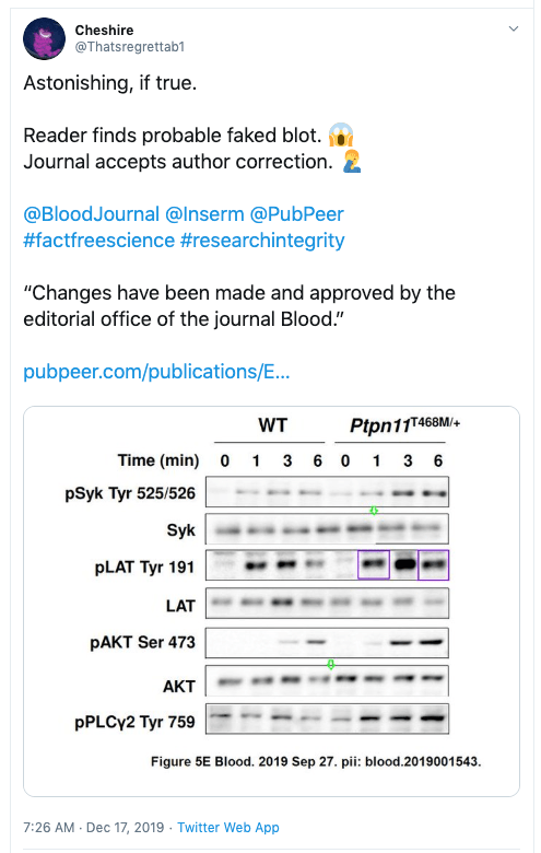
In this case, the authors sort of admitted that something was not right, because they offered a “correction”. Please, @BloodJournal, think critically about accepting a correction to “correct” photo manipulation.
Here is an analogy: If a sports professional had won a race but their blood/urine sample taken just before the race had doping in it, would you accept a clean sample from two weeks later? Would that make it ok? No, it would not.
Unfortunately, many journal editors don’t have the courage to keep science clean. There are dozens of examples where journals rather accept a clean (better photoshopped?) figure redo than asking the authors for a thorough explanation and considering a retraction.
Science has a huge problem: 100s (1000s?) of science papers with obvious photoshops that have been reported, but that are all swept under the proverbial rug, with no action or only an author-friendly correction.
This really has to change. Yes, it is extra work for the journals to adhere to COPE guidelines and contact authors and to show some courage and retract the paper. But dozens of other scientists might try to reproduce these manipulated papers and fail, wasting money, time, sweat, and tears.
Let’s give an “award” to journals that fail to address scientific papers with photoshopped/other obvious problems. Here is my favorite example from the past weeks:

Here is another “weak” decision by the editors of a journal, who accepted a correction to fix several obvious figure manipulations: see my blog post Mega-corrections and weak editors.
The marked images below were spotted by readers and posted on @PubPeer at https://pubpeer.com/publications/05C0E31B381D9E195058AA4CD4FA83 .
Authors reply: “Oops sorry, here is a correction, no results were affected.”
Please, editors @MaterialsToday, show some spine, and retract.
Let me formulate this very clearly:
If a published scientific paper has evidence for significant manipulation in photos or data (duplicated gel lanes, duplicated cells, duplicated parts of flow cytometry plots, etc), a journal should always retract. No correction can ever fix the concerns about the rest of the data.
There, I said it.
I hope @C0PE will agree.
Propose a name for the award
Who can come up with a good name for an award to recognize weak editorial decisions in cases of (what appears to be) significant image or data manipulations? Please reply to this tweet with your suggestions.
@bernardo647: “The CRISPRer”
@Thatsregrettab1: “Yellow duck award”: Yellow signifies lack of courage, Duck signifies shirking responsibility – to which @puddleg replied: And it quacks.
@Nirali01: “Science Fumigation Award”
@BoyceWP: The ‘Inert Invertebrate’ award.
@bradpatrick: Weak T?
@deevybee: The Bik-Brandolini Award – see: https://www.google.com/search?client=firefox-b-d&q=brandolini%27s+law
@MadS100tist: Cold Tea Award
@Leandra2848: The “Ostrich Award”, for hiding from the obvious problems the duplications reflect?
@tarlbot: You may have to own your fame (or infamy) and name the award the “Bik”. I’m sorry you might have to name a bad award for yourself, but if you are the one tipping at those windmills….
@oliphaunt: Weathervane Award?
@ciencia4medica: Pilate prize
@Rikvvz: A. Sayan award, after this glorious incident: https://twitter.com/MicrobiomDigest/status/1194049331638108160
@CC___Raider: Professor X has been awarded “The Floppy” for weakest response to academic misconduct in 2019.


“Let’s give an “award” to journals that fail to address scientific papers with photoshopped/other obvious problems. ”
I nominate Blood.
LikeLike
Blood. 2006 Apr 15;107(8):3295-302. Epub 2005 Dec 27.
NF-kappaB is essential for the progression of KSHV- and EBV-infected lymphomas in vivo.
Keller SA1, Hernandez-Hopkins D, Vider J, Ponomarev V, Hyjek E, Schattner EJ, Cesarman E.
Author information
1
Department of Pathology and Laboratory Medicine, Weill Medical College of Cornell University, 1300 York Ave, New York, NY 10021, USA.
Figure 3A.
https://pubpeer.com/publications/2CF0AD15767F7E22A39DEDC78E40D0#1
LikeLike
Blood. 2007 Jan 15;109(2):729-39. Epub 2006 Sep 7.
Hodgkin lymphoma cells express TACI and BCMA receptors and generate survival and proliferation signals in response to BAFF and APRIL.
Chiu A1, Xu W, He B, Dillon SR, Gross JA, Sievers E, Qiao X, Santini P, Hyjek E, Lee JW, Cesarman E, Chadburn A, Knowles DM, Cerutti A.
Author information
1
Department of Pathology and Laboratory Medicine, Weill Medical College, Cornell University, 1300 York Ave, Rm C-410, New York, NY 10021, USA.
Figure 3B.
https://pubpeer.com/publications/6F8061D70C33EA7D14013FCF2015AB#21
Figures 5A and 5C.
https://pubpeer.com/publications/6F8061D70C33EA7D14013FCF2015AB#17
https://pubpeer.com/publications/6F8061D70C33EA7D14013FCF2015AB#20
LikeLike
Ancient, but does that matter?
Blood. 2000 Feb 1;95(3):1014-22.
Arsenic induces apoptosis of multidrug-resistant human myeloid leukemia cells that express Bcr-Abl or overexpress MDR, MRP, Bcl-2, or Bcl-x(L).
Perkins C1, Kim CN, Fang G, Bhalla KN.
Author information
1
Division of Clinical and Translational Research, Sylvester Comprehensive Cancer Center, University of Miami School of Medicine, Miami, FL 33136, USA.
Figure 3.
https://pubpeer.com/publications/7F63D4DE9AC94C01D9BE1A0680F943#3
Figure 5A.
https://pubpeer.com/publications/7F63D4DE9AC94C01D9BE1A0680F943#4
LikeLike
Blood. 2001 Apr 1;97(7):2031-7.
Signal transduction pathways involved in soluble fractalkine-induced monocytic cell adhesion.
Cambien B1, Pomeranz M, Schmid-Antomarchi H, Millet MA, Breittmayer V, Rossi B, Schmid-Alliana A.
Author information
1
INSERM U364, Facultè de Mèdecine, Nice Cedex 02, France.
https://pubpeer.com/publications/8062A0F364DA11DC293A00C07750C9
LikeLike
Blood. 2001 Jan 15;97(2):359-66.
Signal transduction involved in MCP-1-mediated monocytic transendothelial migration.
Cambien B1, Pomeranz M, Millet MA, Rossi B, Schmid-Alliana A.
Author information
1
INSERM U364, Faculté de Médecine, Nice, France.
https://pubpeer.com/publications/9CA15D90C6CC021393FB7265E9E71A
LikeLike
Blood. 2012 Mar 8;119(10):2335-45. doi: 10.1182/blood-2011-06-361261. Epub 2012 Jan 18.
Targeting of GSK3β promotes imatinib-mediated apoptosis in quiescent CD34+ chronic myeloid leukemia progenitors, preserving normal stem cells.
Reddiconto G1, Toto C, Palamà I, De Leo S, de Luca E, De Matteis S, Dini L, Passerini CG, Di Renzo N, Maffia M, Coluccia AM.
Author information
1
Hematology Unit, Vito Fazzi Hospital Azienda Sanitaria Locale Leece, Italy.
https://pubpeer.com/publications/7D96DF41882229E8ADEFB1B40A7088#4
https://pubpeer.com/publications/7D96DF41882229E8ADEFB1B40A7088#2
LikeLike
Blood. 2005 May 1;105(9):3413-9. Epub 2005 Jan 11.
TRAIL counteracts the proadhesive activity of inflammatory cytokines in endothelial cells by down-modulating CCL8 and CXCL10 chemokine expression and release.
Secchiero P1, Corallini F, di Iasio MG, Gonelli A, Barbarotto E, Zauli G.
Author information
1
Department of Morphology and Embryology, Human Anatomy Section, University of Ferrara, Via Fossato di Mortara 66, 44100 Ferrara, Italy.
Figure 1C.
https://pubpeer.com/publications/84595DDA1238D638929B782D35B854#3
LikeLike
Blood. 2006 Jan 15;107(2):508-13. Epub 2005 Sep 15.
PKCepsilon controls protection against TRAIL in erythroid progenitors.
Mirandola P1, Gobbi G, Ponti C, Sponzilli I, Cocco L, Vitale M.
Author information
1
Department of Anatomy, Pharmacology, & Forensic Medicine, Human Anatomy Section, University of Parma, Ospedale Maggiore, Via Gramsci, 14, I-43100 Parma, Italy.
Figure 2A.
https://pubpeer.com/publications/FE55D68ED423DC39FEE759C9BD9617#3
Figure 6B.
https://pubpeer.com/publications/FE55D68ED423DC39FEE759C9BD9617#2
LikeLike
Blood. 2006 Jan 15;107(2):514-9. Epub 2005 Oct 6.
Von Hippel-Lindau-dependent polycythemia is endemic on the island of Ischia: identification of a novel cluster.
Perrotta S1, Nobili B, Ferraro M, Migliaccio C, Borriello A, Cucciolla V, Martinelli V, Rossi F, Punzo F, Cirillo P, Parisi G, Zappia V, Rotoli B, Della Ragione F.
Author information
1
Department of Pediatrics, Second University of Naples, Via Luigi De Crecchio, 4, Naples, Italy.
Figure 2B.
https://pubpeer.com/publications/3C2F388723490D3EB719ABD44FEF9B#2
Figures 2B and 3B. Figure 3A.
https://pubpeer.com/publications/3C2F388723490D3EB719ABD44FEF9B#3
Figures 5A and 5B.
https://pubpeer.com/publications/3C2F388723490D3EB719ABD44FEF9B#5
LikeLike
Elisabeth m bik!You take bribes from researchers in USA. You are just a whore who aims at making money!
LikeLike