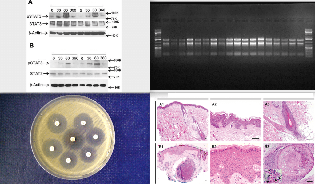
One of my recent investigations led me to expand my set of figure types to look at. For our 2016 mBio study, in which I scanned >20,000 papers for image duplication, I focused on real photos of Western blots, agarose gels, tissue sections, etc.
One of my recent investigations led me to expand my set of figure types to look at. For our 2016 mBio study, in which I scanned >20,000 papers for image duplication, I focused on real photos of Western blots, agarose gels, tissue sections, etc.

