This week I worked on a large set of papers from a research group at the Tianjin Life Science Research Center at Tianjin Medical University. The group, headed by Dr. Hua Tang and funded by many National Natural Science Foundation of China grants, has published a total of 113 PubMed-indexed papers.
However, a significant number of these — 45 as of today — have PubPeer posts in which concerns are raised about their figures.

Cancer research
Tianjin Medical University (TMU), founded in 1951, is the oldest medical institution approved by the State Council of the People’s Republic of China. TMU’s Cancer Institute and Hospital (TMUCIH) is one of the largest cancer centers in China.
Not surprisingly, many research groups at TMU study cancer as well. One such group is Dr. Hua Tang’s at TMU’s Tianjin Life Science Research Center. Their research focuses on the role of microRNAs (miRNAs) in the development of several types of cancer, such as liver carcinoma, gastric cancer, and cervical cancer.
Measuring the growth of cancer cells
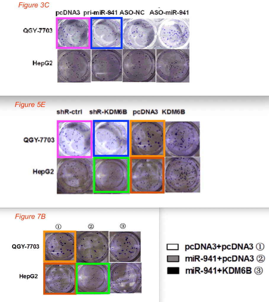
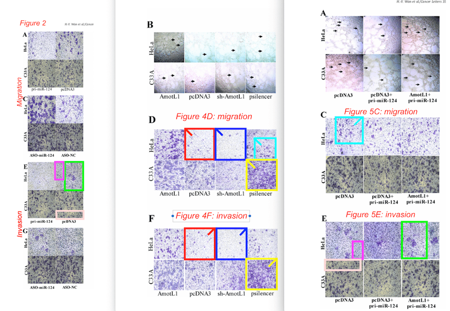
The papers from the Hua Tang group often include multiple panels with microscopy photos of cancer cells grown with or without certain miRNAs, to see how this affects the cells’ ability to migrate, invade, or form colonies in soft-agar. Simply put, a photo of an experiment showing many cells means the cancer cells are growing abundantly, while fewer cells in a photo mean that the cancer cells are inhibited. Figures in these papers contain photos of the differently treated cells taken under a microscope (shown at the bottom in the example below), and the measured number of cells in each experiment (shown at the top).

Similar and overlapping panels
While the example above raises no image concerns on its own, some of its panels appear to overlap with panels in other figures from the same paper.
Here is a comparison of several figures from the same paper, with colored boxes showing very similar areas found by PubPeer users Actinopolyspora Biskrensis, Nymphalis Xanthomela (both pseudonyms), and me:
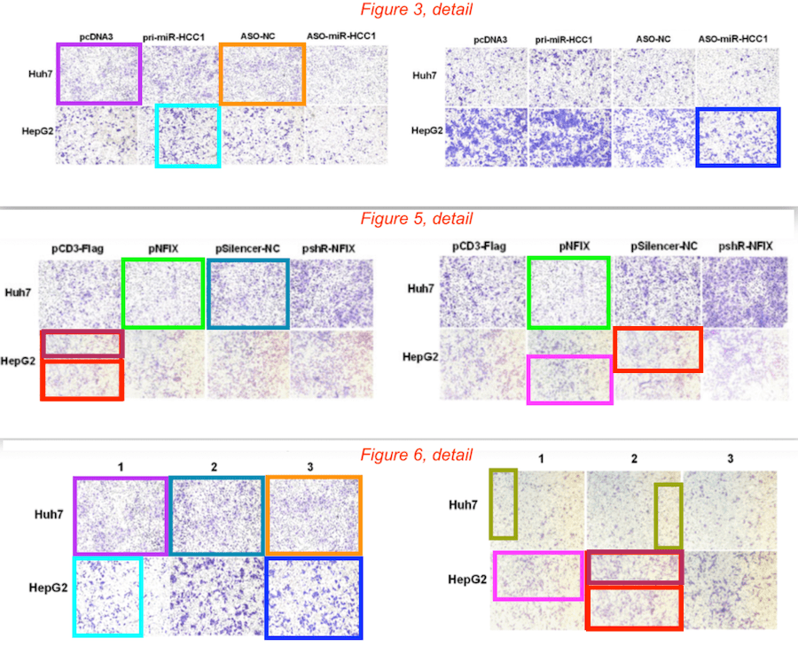
Many of Tang’s papers raise similar concerns. Here is a collage of three figures from another paper, in which I have highlighted colony-formation wells that appear to look very similar.
Another paper with similar concerns is shown below. Some panels in this paper seem to have been rotated, which I’ve indicated using a diagonal stripe in one of the corners. In other cases part of the panel appears to be visible multiple times. These areas of overlap suggest that the same sample was photographed more than once from different angles, then used to represent different experiments.
Forty five papers and counting
As of today, the Tang research group has 45 papers flagged on PubPeer. If you do this search, the number of PubPeer posts appears a bit higher, but some posts are about papers with other authors with a similar name, and the site also shows some entries multiple times.
One of the papers from this group, Xue Liu et al., PLOS ONE (2013), DOI: 10.1371/journal.pone.0064707 was found during my study of 20,000 papers back in 2014/2015. I reported it to the journal in October 2015, and it was retracted in May 2020, almost five years later. You can take a look at the image concerns on PubPeer or in the retraction notice.
For now, I have reported the other papers to the editors of the journals in which they were published. Let’s hope an update will not take another five years.
Here are the 45 papers:.


Hi great reading your blogg
LikeLike