
As I was doing a reverse-image search online on a set of papers, I saw an unrelated photo of a skin biopsy that appeared to contain repetitive areas. Even in the small Google Image Search thumbnail it was clear to me that something unexpected was going on in this image.
The discovery led me to a set of 12 papers from a research group at the Hematopathology Department of the Texas MD Anderson Cancer Center, all with histopathology photos showing unexpected repetitive structures.

The Magic Eye picture
The paper in question — Seervai et al., J Cutan Pathol (2022), DOI: 10.1111/cup.14145 — was written by authors affiliated with Baylor College of Medicine, and the University of Texas MD Anderson Cancer Center.
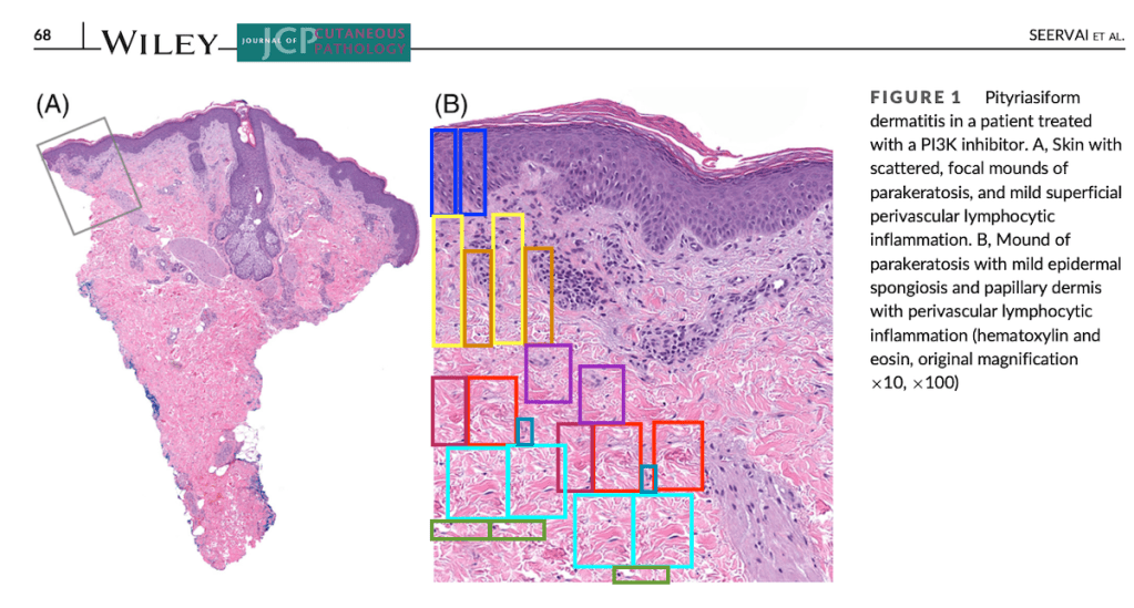
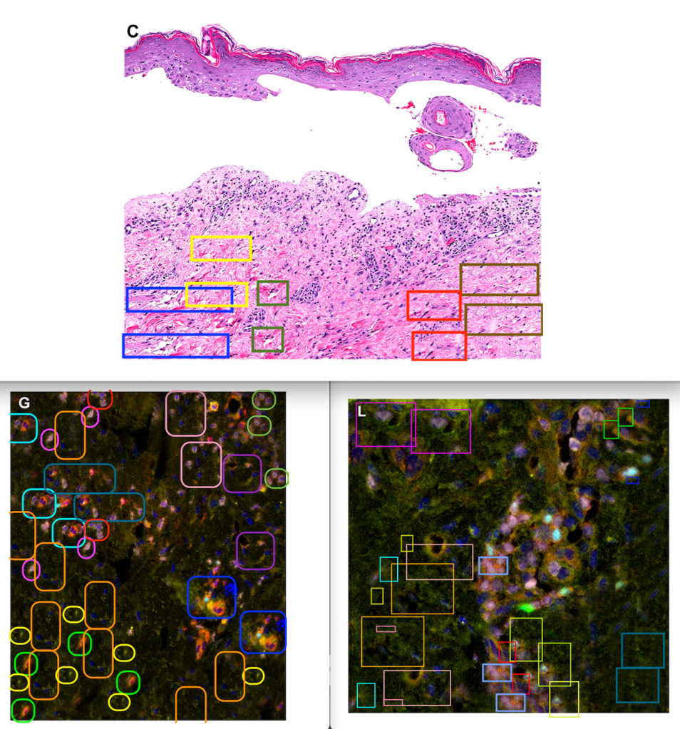
The skin biopsy image that I spotted in Figure 1 had a lot of repetitive features in the bottom half of the photo. In fact it looked like one of those Magic Eye autostereogram photos that were so popular in the 1990s.

I stared at the skin image for a while, hoping I might see a dolphin or a pyramid, but nothing 3D popped out. A bit disappointed, I started marking the image with boxes of the same color to highlight areas that looked similar. And there were a lot!
Most duplicated areas in Figure 1B were in the bottom left corner. Comparing panel B to lower-magnification panel 1A, it appeared that a big empty area with no tissue had been digitally filled-in with content from elsewhere in the photo.
DOI: 10.1111/cup.14145. See PubPeer: https://pubpeer.com/publications/B52B2B4800FDA6B38FC24AEBB96671
More figures-of-concern in the same paper
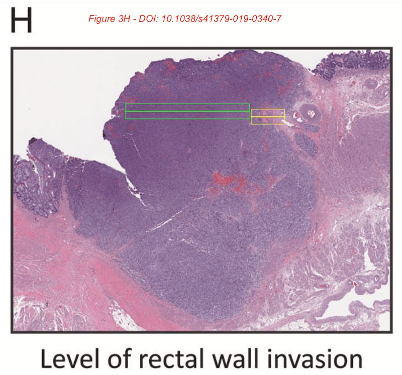
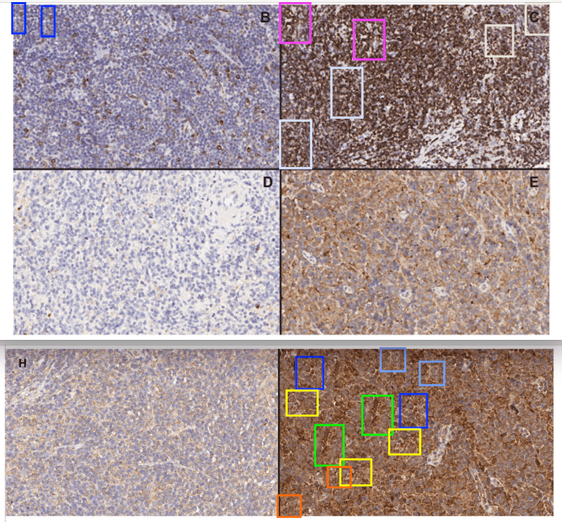
Figures 2 to 4 from the same paper also appeared to contain repetitive areas in the high-magnification panels. I marked them all with boxes of the same color and posted my concerns on PubPeer.

Surprisingly, this was a review paper. It’s unusual for review papers to contain photos, but when they do there’s usually an acknowledgment that the figures were reproduced from another paper. However this paper contained no wording indicating that the figures came from other studies. Confusingly, the paper also stated that ‘no data are available’.

So were the photos in Figures 1-4 not data? Did these patients really exist? I had so many questions. Unfortunately, so far none of the authors have replied to the questions I posted on PubPeer.
Corner cloning and stitching mishaps
Investigating more papers from the same researchers yielded an additional 11 papers [PDF list here: md-anderson-pathology-papers-google-sheets] containing histopathology photos with similar repetitive areas. In some cases the problems were relatively benign.
In one instance, the repetitive areas might have been caused by a mishap in ‘stitching’ together two images to produce a large overview photo of a piece of tissue. This would be similar to stitching errors in panorama or 360 degree photos — the software can get confused, possibly leading to some repetitive areas.
In another paper, DOI: 10.1136/jclinpath-2018-205417, three figures all had duplicated areas in the bottom left corner. I call this “Corner Cloning“, where one area close to the corner of a photo contains a repeat seen elsewhere in the photo, but where no other repeats are found. This might be to cover up a scale bar or some imprint added by the microscope software.
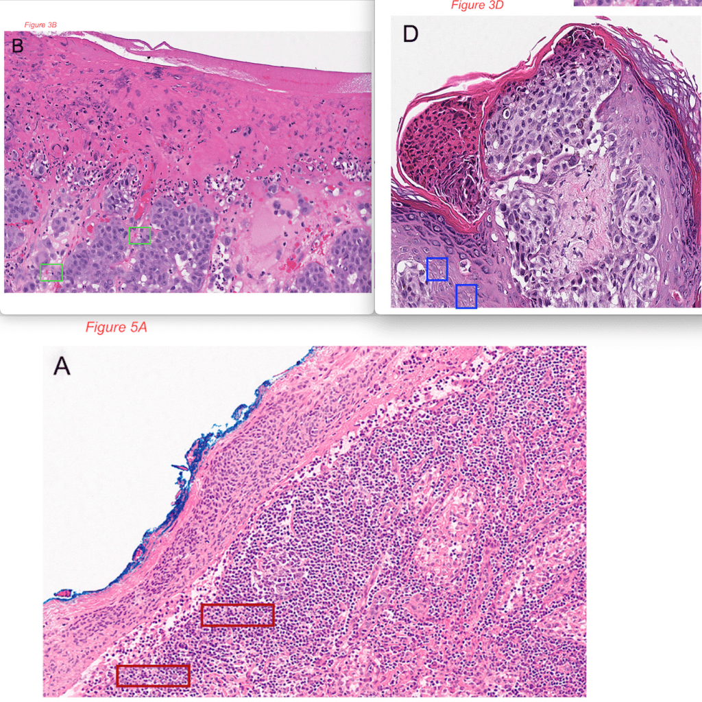
For example in this 2020 Histopathology paper about Kikuchi-Fujimoto Disease, small corner clones were found in three panels of Figure 4.
More serious concerns in MD Anderson papers
In other cases, the duplications appeared to be rather more concerning. This 2020 paper about Epstein Barr Virus, published in the journal Pathology, contains at least three figure panels with lots of duplicated areas.

Another Epstein Barr Virus paper published in Modern Pathology (2021) also contained a photo that appears to have been digitally altered.

A 2021 paper published in Frontiers in Oncology about urothelial carcinoma treatment showed extensive duplication of areas in panels in Figures 2 and 4. In addition the case reports described in the study did not have informed consent or permission-to-publish from the patients. And in a severe breach of peer-reviewers’ code of conduct, both peer reviewers were recent collaborators with the authors on other studies.
PubPeer comments with author replies
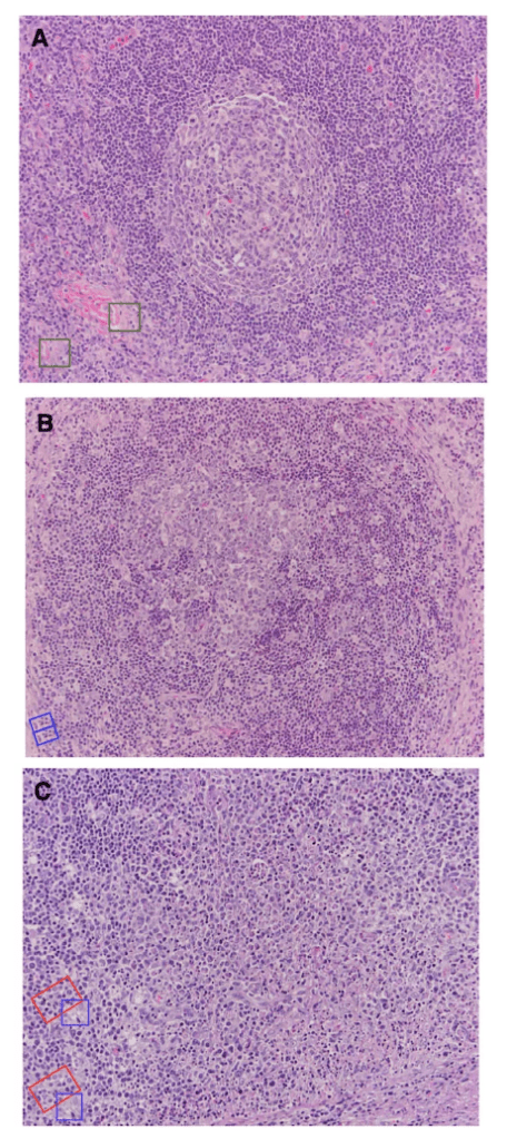
A paper about lymphomas published in Blood Cancer Journal (2021) also raised concerns. Figure 1 appeared to contain multiple repetitive areas (shown below), while some corner cloning is apparent in a supplemental figure. On PubPeer the first author replied that a scale bar had been removed from the supplemental figure, but said that the Figure 1 photos had not been changed. He provided high-resolution versions of these panels, but unfortunately they showed even more duplicated areas.
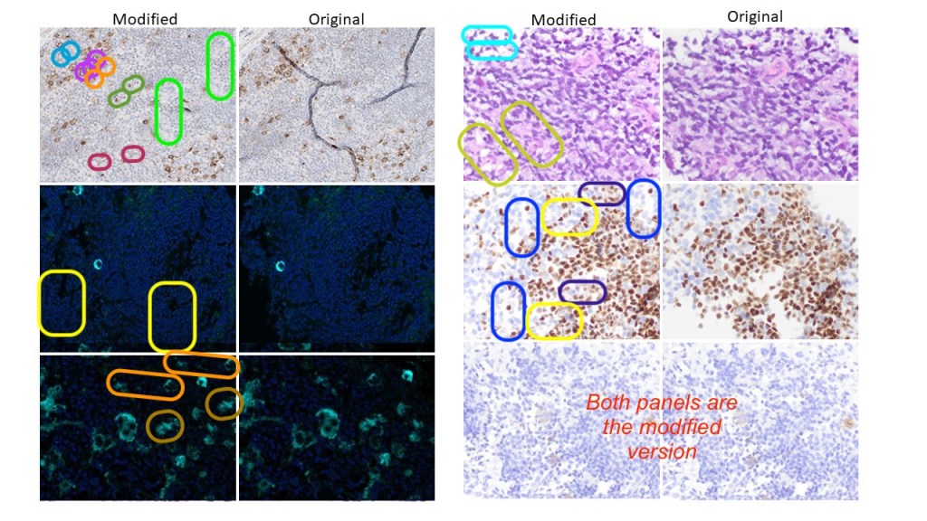
In this Leukemia & Lymphoma (2021) paper, Figure 2 was found to contain duplicated areas in two of its panels. In this case the senior author replied on PubPeer by posting the original photos and explaining that ‘minor improvements’ were made to fill up holes in the tissue. This is like filling up the holes in Swiss cheese with more cheese.

Another PubPeer author-reply and a set of original, unaltered photos were received for this Histopathology (2023) paper about Hodgkin lymphoma, with six panels containing repetitive areas. The senior author admitted to “slightly modifying images to improve their visual appeal”. Comparing published vs. original photos revealed that the cloning was performed to fill holes, remove some black vein-like structures, or add more cells. The photos might now have improved visual appeal, but one can only wonder how scientific and truthful the published results actually are.
Conclusions
A set of 12 papers containing repetitive areas in histopathology photos was discussed above. These papers all shared affiliation with the University of Texas MD Anderson Cancer Center’s Departments of Hematopathology or Pathology. Although there wasn’t one common author across the 12 papers, some author names occurred multiple times, such as Marques-Piubelli ML (8 papers) and Miranda RN (5 papers).
The problems found in these figures ranged from mild (stitching errors or corner cloning to remove a scale bar) to serious — where gaps in the tissues appear to have been filled up with other parts of tissues. Most duplications were confirmed by ImageTwin.
Today, most journals have image guidelines that specifically prohibit such alterations of photos. And since most of these papers are less than 3 years old, such guidelines should have been known to the authors and followed.
You could argue that ‘gap filling’ (filling up the holes in Swiss cheese with more cheese) would be akin to filling up missing data in a spreadsheet with data found in other rows — something that would clearly be viewed as data fabrication. But maybe this sort of ‘beautification’ of photos is quite accepted among pathologists? Do let me know your thoughts in the comments.
A list with all 12 papers [PDF] can be found below. I’ll update the list if I find more problems in this set.


I am a pathologist. When writing papers, I would never alter an image in the ways that you are suggesting. If there were a tear in the section at a crucial spot, I would leave it. Other pathologists are perfectly capable of mentally stitching things together.
I also would never have picked up on the duplications that you are showing. I don’t think that I have the skills.
I wonder how common this is in the pathology literature.
LikeLike
Thank you for highlighting this. At Histopathology we have clear guidelines for what is acceptable and what is not acceptable. As editor in chief I will be taking this forward: we do have image manipulation software and I will be investigating to find out whether this was detected and the next stages we need to take.
Dan Berney, Editor in chief Histopathology
LikeLike
This is very common in the scientific world. The pressure to publish makes people do unethical things.
LikeLike
What was the outcome of MDS’s investigation into the allegations? I assumed you contacted them first as to keep the matter confidential and protect evidence/data before going public. When internet vigilantes publicly post allegations of research misconduct before an institution has had adequate time time to sequester the data, the data suddenly goes missing, often significantly thwarting the investigative process and unfairly publicly accusing the author(s) of career damaging allegations of research misconduct before any investigation has occurred.
LikeLike
I’m late to this, but “image (B)” does work as a Magic Eye picture! Focusing on the adjacent areas you outline in red in your analysis and allowing those structures to overlap gives a fine 3D image of floating planes above a flat background.
LikeLike