This blog post expresses my personal opinion and is not an accusation of misconduct.
An exciting new paper about STAT3
The STAT (“signal transducer and activator of transcription“) protein family consists of proteins involved in many important aspects of cellular function, such as growth, differentiation, and apoptosis (programmed cell death). These transcription activators are activated themselves if other molecules bind to them, and they act as messengers that transfer changes outside of a cell to inside the nucleus, by binding to promoters and determining which genes are switched on or off. One of STAT proteins, STAT3, in particular has been the topic of many studies, because it might play a role in cancer. Simply put, the continuous activation of STAT3 might induce cancer, and STAT3 might be a target for new anti-cancer drugs.
A recent study, published on 28 August 2019 in Nature by authors from Harvard Medical School and the Dana Farber Cancer Institute, therefore gained quite some attention. It reported on one of the ways by which STAT3 can be activated, through the binding of fatty acids in a process called palmitoylation.

The paper included four figures, each consisting of several smaller panels. Many of these were Western blots, photos of proteins stained by specific antibodies to study their expression in different experimental conditions.
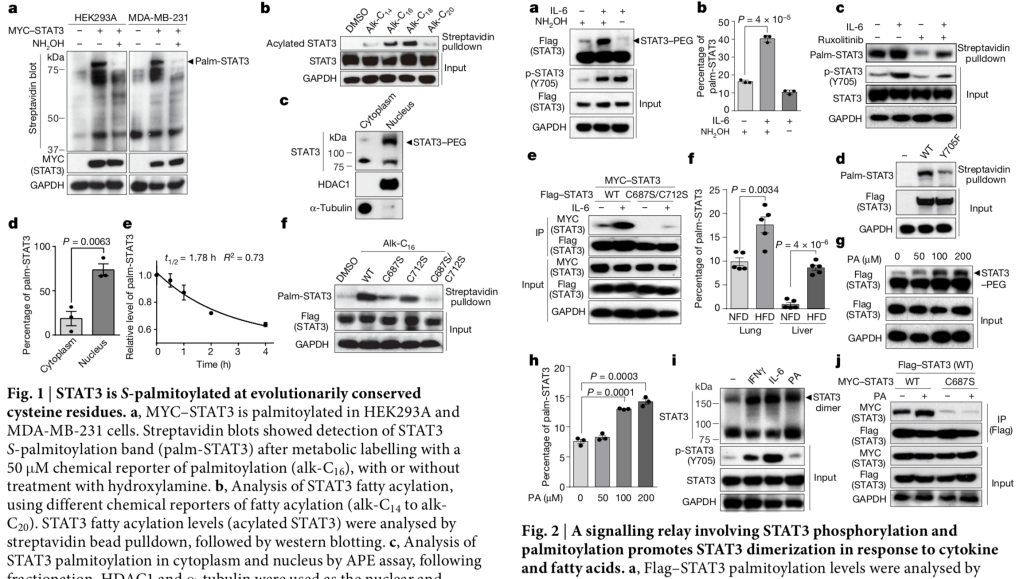
All bands in a Western blot (the black horizontal thick “stripes”) should be unique. Like faces, rocks, clouds, or snow flakes, each band and surrounding area will look different than the other bands, and one would never expect to see the same band twice within the same photo. In other words, two bands that represent different experiments should never look the same.
Because I have screened so many papers in the past for image duplication, I could not help but looking at the Western blot photos within this Nature paper. Within seconds, I noticed that something was not right.
Unexpected image similarities
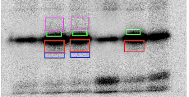
Although the bands had different shapes, the areas just above and below some bands appeared to be repeated in other lanes within the same photo. Here is a figure illustrating the patterns I was seeing in three figure panels in Figures 2 and 3. The colored boxes were added by me, and boxes of the same color highlight areas that look unexpectedly similar to each other.
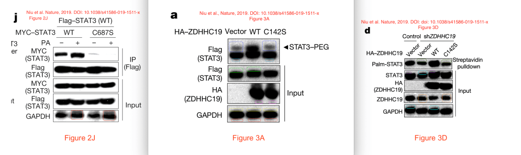
This might be very hard to see, so let’s zoom in. Here is the GAPDH panel of Figure 2J in more detail. Red and burgundy boxes highlight two areas above and below two bands that look unexpectedly similar. Note that the bands have different shapes, and the areas differ in brightness, but the pattern of lighter and darker dots in these areas look very similar to me.

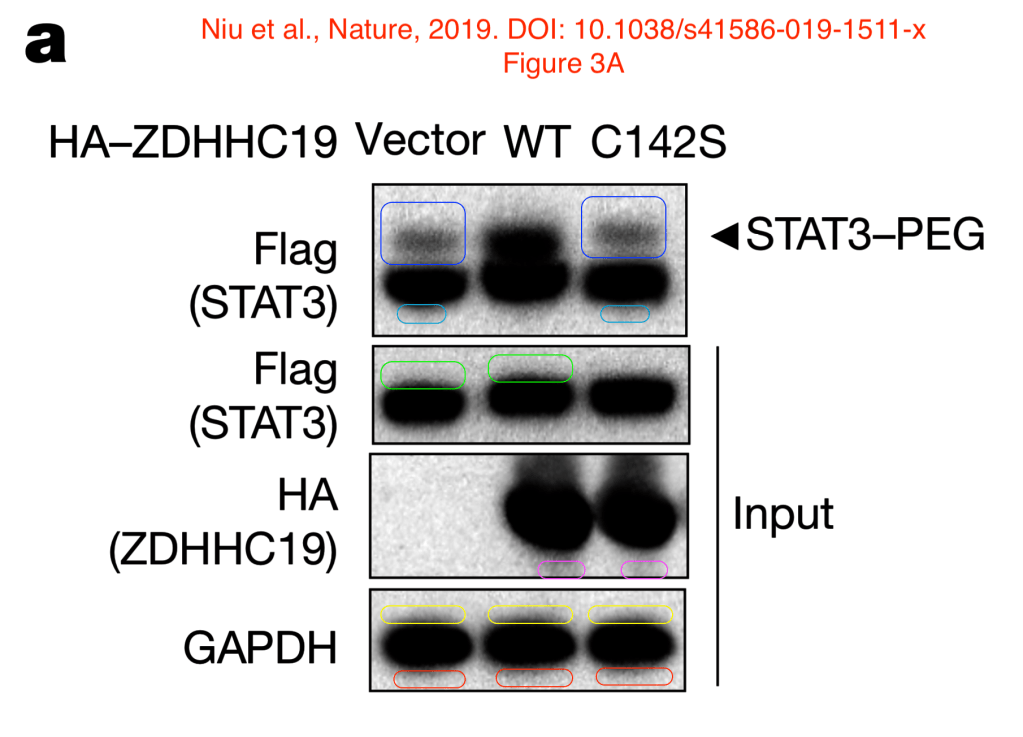
All panels in Figure 3A appear to have such similarities as well. Here is a zoomed-in version.
Finally, here is a close-up view of Figure 3D. Of note, the same area appears to be visible once in the STAT3 panel and twice in the GAPDH panel (shown in aqua blue boxes).

In each of these cases, the bands themselves appear to be different in width, thickness, and shapes, but the surrounding areas are similar. So this is not a case where one lane was simply duplicated to represent a different experiment.
The Nature paper provided uncropped scans of these blots, but they appeared to have the same duplications, so they did not help in decreasing my concerns about this paper.
Reporting my concerns
I reported my findings to Nature, the journal in which this paper was published. I also wrote about this on Twitter and on PubPeer. This all happened on August 29, a day after the paper had been published online.
Nature immediately replied both on Twitter as well as per email that they would look into this. This has been two months ago, but I have not heard any updates.

Similar problems in other papers
Meantime, I searched for other papers by the same authors to see if they had similar image problems. Other papers from the last author on the Nature paper were all clean, but several older papers by the first author on the Nature paper appear to have similar image duplications.
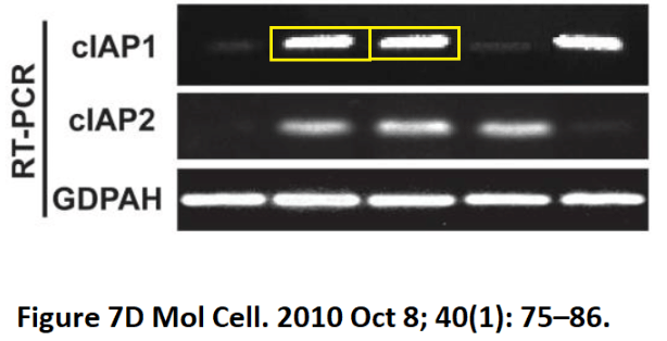
Zhao-Hui Wu et al., Molecular Cell (2010) had already been flagged on Pubpeer by “Cyathea Longipinnata” (pseudonym), for a possible duplicated band in Figure 7.
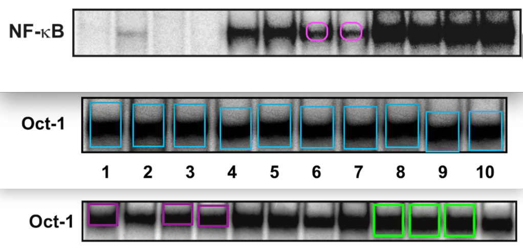
Jixiao Niu et al., EMBO Journal (2011) appears to have duplicated lanes in several figures, as highlighted on PubPeer. Here is a selection of some of these similarities, all highlighted with colored boxes.
Jixiao Niu et al., Journal of Biological Chemistry (2012) has several possible problems as well, and the PubPeer discussion is ongoing.
The senior author posted the “original scan”, but it did not take away any concerns. As pointed out by Hoya Camphorifolia and confirmed by Forensically, the original scan still appeared to have the duplication, and another one as well.
Finally, a fourth paper, Jixiao Niu et al., EMBO Journal (2013), had possible duplications in three of its figures, as discussed on PubPeer.

Investigations ongoing
Both Nature and EMBO Journal were quick to reply that they would investigate these concerns. These types of high-profile papers might take months to investigate, and it is fair to give the authors a chance to come up with original scans or explanations.
It took Nature about 6 months to retract the Obokata papers, after concerns were raised on social media.
I will update this post as soon as I have more information.


This is very problematic clearly and Nature should act swiftly to avoid the case escalates.
LikeLike